Coccidiosis in Cattle
Quick Facts
The Impact of Coccidiosis
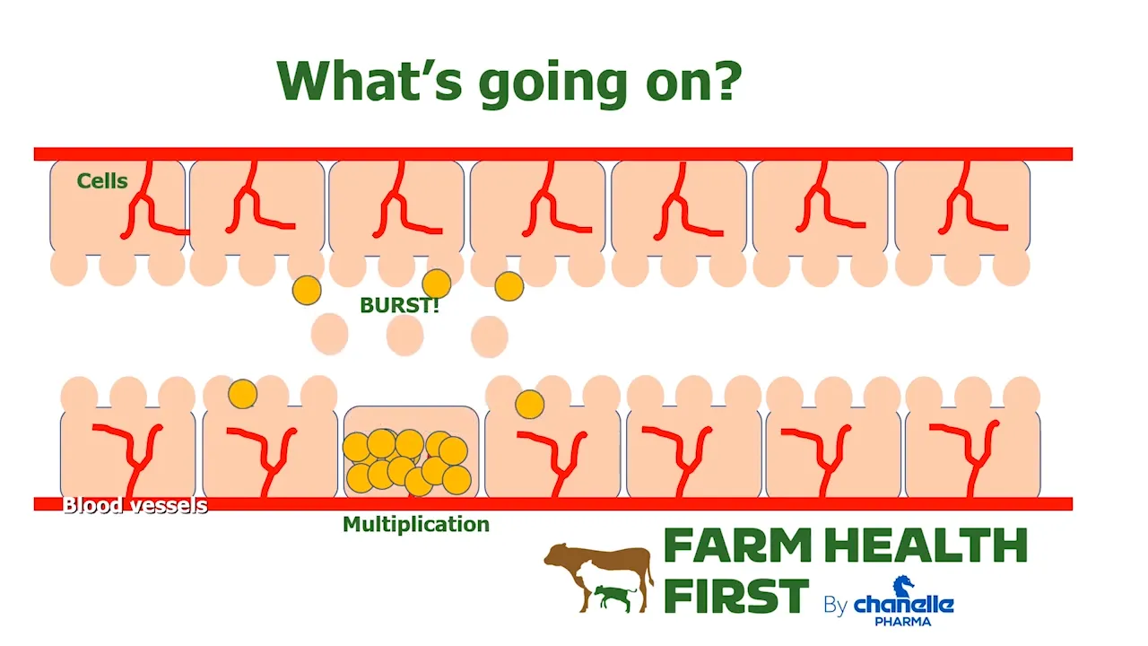
Coccidiosis is a common and costly disease of young livestock, including calves. It damages the lining of the gut, reducing nutrient absorption and growth.
Clinical disease can cause:
- Scour (sometimes with blood)
- Ill thrift and poor growth
- Increased susceptibility to other diseases
- In severe cases, death
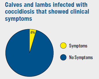
However, subclinical disease is much more common and causes the most financial damage to a farm.
What causes coccidiosis?
Coccidiosis is caused by coccidia, microscopic protozoal parasites, which all ruminants kept under normal conditions will encounter.1,2
The main pathogenic species in calves are:
- Eimeria bovis
- Eimeria zuernii
A third species, Eimeria alabamensis, is associated with pasture-based disease after turnout:
- Disease often seen 1–3 weeks after turnout
- Short pre-patent period (6–8 days), so disease can develop quickly following exposure
- Causes a noticeable growth check but rarely fatal
It’s important to remember:
- Coccidia are host-specific – cattle coccidia do not infect sheep, and vice versa
- Many non-pathogenic coccidia species can be found in healthy animals without causing disease
- Disease occurs when pathogenic species are present in high numbers, particularly in young or stressed animals
- Coccidia oocysts (eggs) are highly resistant, surviving for long periods in housing and at pasture, including overwinter. They also thrive in damp conditions.
Understanding how coccidiosis spreads, which animals are most at risk, and how to manage housing, grazing and treatment strategies is key to reducing losses and improving growth rates.
How Does Coccidiosis Spread?
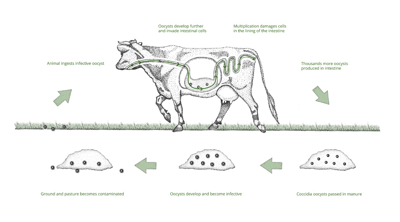
Calves become infected by ingesting coccidia oocysts while exploring their surroundings. Due to the rapid multiplication of coccidia, later-born animals are most at risk.
Most coccidia species, except Eimeria alabamensis, have a pre-patent period of approximately 21 days. This is the time between ingestion of the parasite and shedding of new oocysts in the dung.
Immunity and Disease Risk
Under natural conditions, all young animals are exposed to low levels of coccidia. This causes only minor gut damage, which is easily repaired, and the exposure helps develop immunity.
However, disease can develop rapidly when:
- Environmental contamination with pathogenic coccidia species is high.
- Naïve animals, often immunocompromised due to age or other stressors, are exposed.
Clinical Signs in Calves
Disease is most commonly seen in calves between 3 weeks and 9 months of age, depending on the production system and level of exposure.
Acute Disease
Typically, only some animals within a group are acutely affected.
- Sudden onset of profuse scour
- Straining may be present
- Blood may be present in the dung
- Rapid dehydration
- Inappetence
- Depression and weakness
Chronic Disease
Often the whole group is chronically exposed to high parasite burdens.
- Calves begin "losing their shine"
- Often the first sign of disease within a group, but can easily be overlooked because it is non-specific
- Paste-like grey dung
- Reduced appetite or enthusiasm
- Ill thrift and poor growth
- Increased susceptibility to other diseases due to immune suppression
- Chronically stunted animals
System-Specific Patterns
Dairy calves and milk-fed calves
- Typically affected between 3 and 12 weeks of age.
- Disease is associated with the build-up of contamination within calf housing.
Suckler calves
- Disease can occur both indoors and at pasture.
- Higher risk exists around contaminated water sources, troughs and poached areas.
- As oocysts survive on pasture, fields grazed by calves year after year carry an increasing disease challenge.

Diagnosing Coccidiosis in Calves
Diagnosis requires careful interpretation, and it is always best to seek veterinary advice.
Faecal Sampling
- The most useful rapid diagnostic tool.
- Sample several calves within the group, not just one sick animal.
- Clinical signs may occur before oocyst shedding begins.
- The presence of coccidia alone is not diagnostic, as many non-pathogenic coccidia species are commonly found in healthy animals.
High counts of a single pathogenic species, alongside compatible clinical signs, are suggestive of coccidiosis.
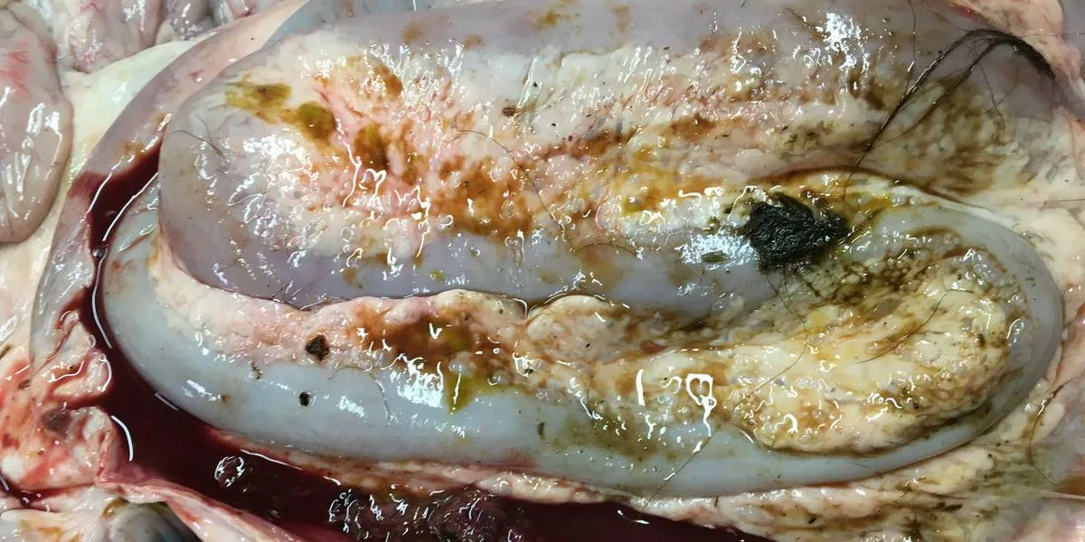
Post-mortem Examination
- Useful in cases of acute deaths to rule diseases in or out.
- It can be more difficult to establish the original cause in chronically poor-doing calves.
Preventing Coccidiosis in Calves
Good control relies on reducing environmental contamination while protecting susceptible animals.
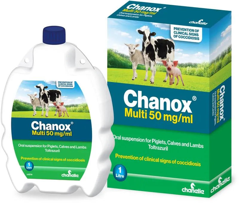
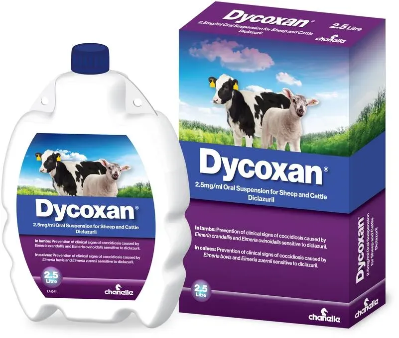
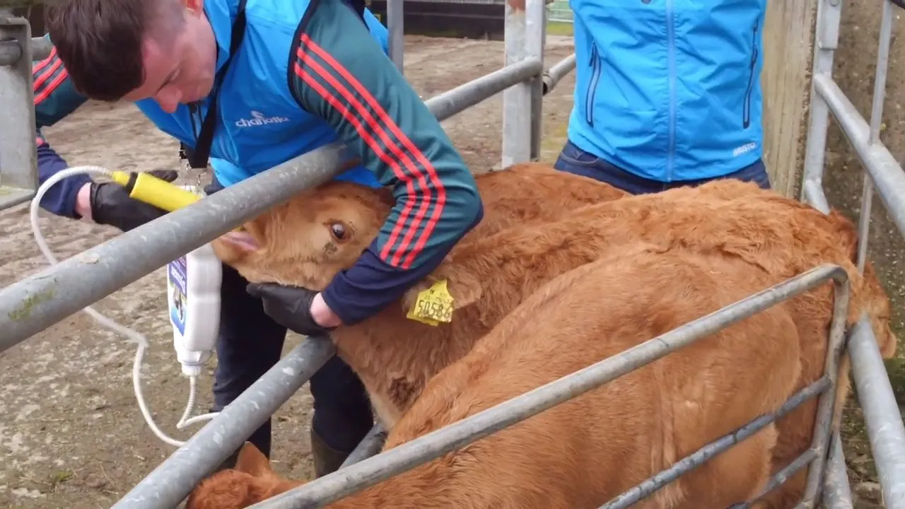
Speak to your vet about timely treatment with either diclazuril or toltrazuril as a routine prophylactic measure to help prevent disease and reduce environmental contamination on your farm.
Coccidiostats can also be added to feed under veterinary prescription as an alternative control measure. This approach relies on all youngstock consuming a consistent amount of feed each day but does not allow animals to develop their own immunity through natural exposure.
Managing a Disease Outbreak
Before treating, ensure coccidiosis is the most likely cause, as many conditions can cause scour and ill thrift in calves. If your farm has a known history of coccidiosis, treatment should begin as soon as signs are observed.
Treatments such as diclazuril and toltrazuril should always be used alongside appropriate management and control measures.
Supportive Care
Under veterinary guidance:
- Rehydration and electrolyte therapy tailored to the individual calf.
- NSAIDs for sick animals where appropriate.
Environmental Control
- Thoroughly clean and disinfect pens, water troughs and feeders.
- Check that the disinfectant used is effective against Eimeria species.
- Isolate scouring animals.
- If calves are at pasture, move them to clean grazing where possible.
References
- Daugschies, A., et al. (2007). Veterinary Parasitology, 149, 199-206.
- Platzer, B., et al. (2005). Veterinary Parasitology, 129, 1-9.
- Romero, J., et al. (2013). Veterinary Parasitology, 193, 277-280.
- Scala, A., et al. (2014). Small Ruminant Research, 120, 242-246.
- Enemark, H. L., et al. (2015). Parasitology Research, 114, 201-212.
- Mundt, H. C., et al. (2009). Parasitology Research, 105, 141-150.
- Diaferia, M., et al. (2013). Parasitology Research, 112, 163-168.
Products

Videos




















Blog

.webp)
.webp)
.webp)
.webp)
.webp)
.webp)
.webp)
.webp)







%2520-%2520What%2520is%2520coccidiosis_%25200-27%2520screenshot.webp)

More Diseases
Know the solution.


CPD for SQPs & Vets
Sharpen your knowledge

DOsing Best Practices
Get the dose right every time