Liver Fluke in Cattle
Quick Facts
The Impact of Liver Fluke
Liver fluke is a parasitic disease caused by Fasciola hepatica. It is one of the most economically important parasites affecting grazing livestock across the UK and Ireland.
The parasite damages the liver as immature fluke migrate through the organ before later feeding within the bile ducts.
Losses can occur through:
- Poor feed conversion
- Reduced growth rates
- Reduced milk yield
- Liver condemnation at slaughter
- Increased susceptibility to other diseases
Fluke infection is strongly influenced by weather, grazing conditions and the presence of mud snails, which act as the parasite's intermediate host.
Economic Impact
In cattle, liver fluke infection can reduce:
- Meat production by up to 20%
- Milk production by up to 8%
How Does Infection Occur?
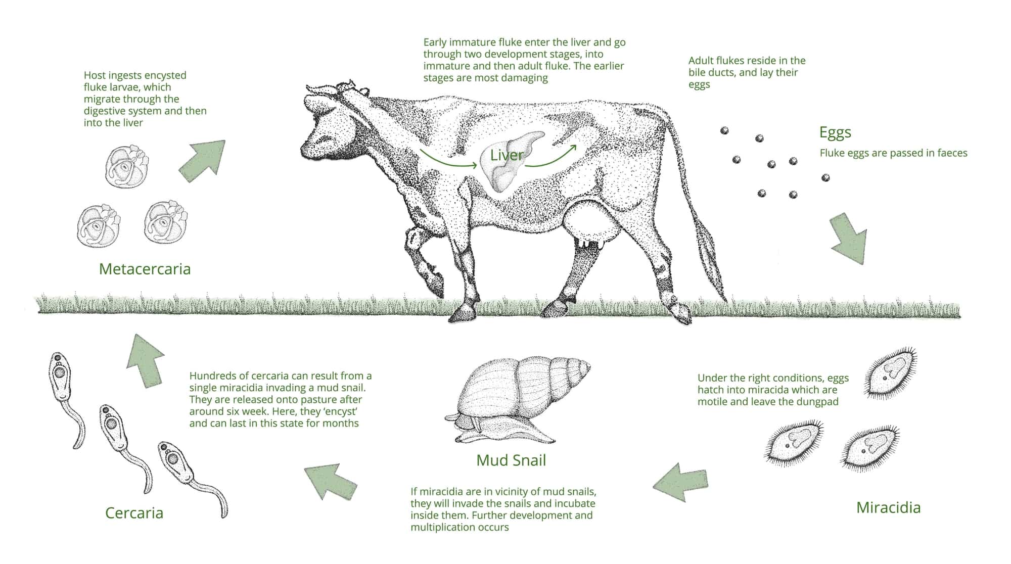
The lifecycle of Fasciola hepatica requires a mud snail as an intermediate host, where larvae continue their development. The encysted larvae (metacercariae) can survive on pasture for many months.
What Impacts Fluke Risk?
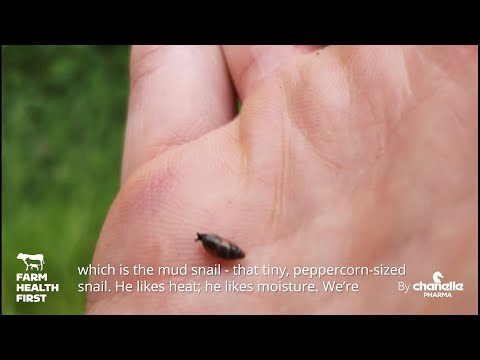
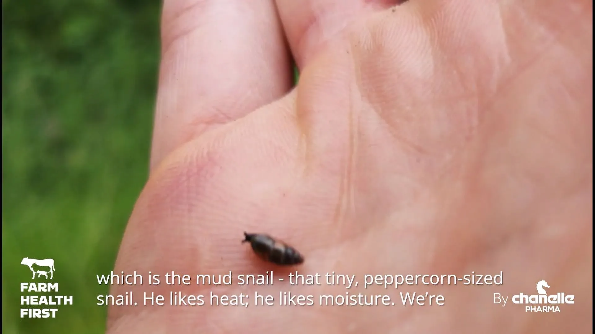
Fluke transmission depends on the mud snail (Galba truncatula). These snails thrive in wet, boggy ground, making poorly drained fields, muddy gateways and water trough areas fluke hotspots.
Warm, wet summers and mild winters increase both snail populations and parasite development, which is why fluke risk is increasing with changing weather patterns.
First-season grazing spring-born calves, especially beef sucklers, are usually low risk early in the grazing season. However, risk increases later in the year as pasture contamination develops. Dairy calves may also be exposed if turned out onto heavily contaminated pasture.
It is important to remember that larvae can overwinter within the mud snail and contaminate grazing the following season.
Sheep are often responsible for introducing and maintaining liver fluke on beef and dairy farms. Tack sheep, particularly those coming from higher-risk farms, can represent a significant source of infection.
Wildlife such as deer and rabbits can also act as hosts, meaning pasture contamination may continue even where livestock have not recently grazed.
Clinical Signs of Liver Fluke in Cattle
Cattle usually develop chronic disease, which may result in significant production losses without obvious clinical illness.
Signs include:
- Reduced weight gain
- Reduced milk yield
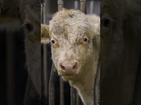
- Bottle jaw (fluid swelling beneath the jaw)
- Pale mucous membranes (anaemia)
- Poor body condition
Because these signs are often subtle, infections frequently go unnoticed until animal performance declines.
Unlike sheep, cattle do not normally suffer acute disease caused by immature liver fluke.
Diagnosing Liver Fluke
Diagnosis may involve several tests.
Faecal Egg Counts (FECs)
- Detection of eggs confirms adult liver fluke infection.
- Eggs generally only appear 10 to 12 weeks after infection.
Blood or Milk ELISA
- Detect antibodies against liver fluke.
- Can identify exposure earlier than faecal egg counts in young animals.
- Interpretation is more limited in older animals because it is difficult to determine when exposure occurred.
Coproantigen Testing
- Detects liver fluke antigens in faeces before egg production begins.
Your vet will combine test results with farm history and seasonal risk when diagnosing liver fluke infection.


Flukicide Treatments
Several active ingredients are available for controlling liver fluke:
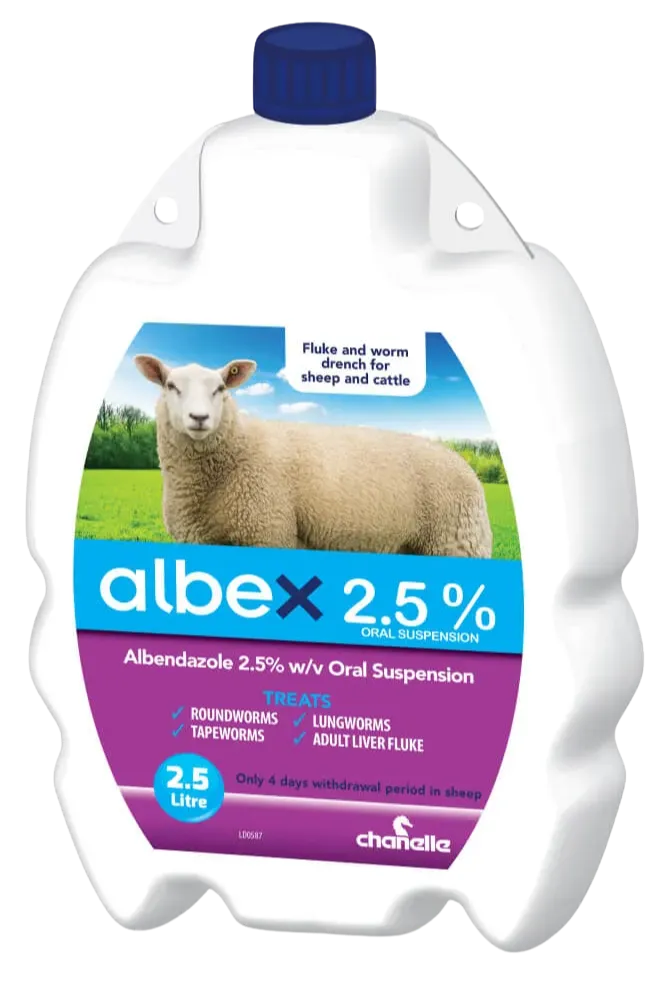
- Albendazole
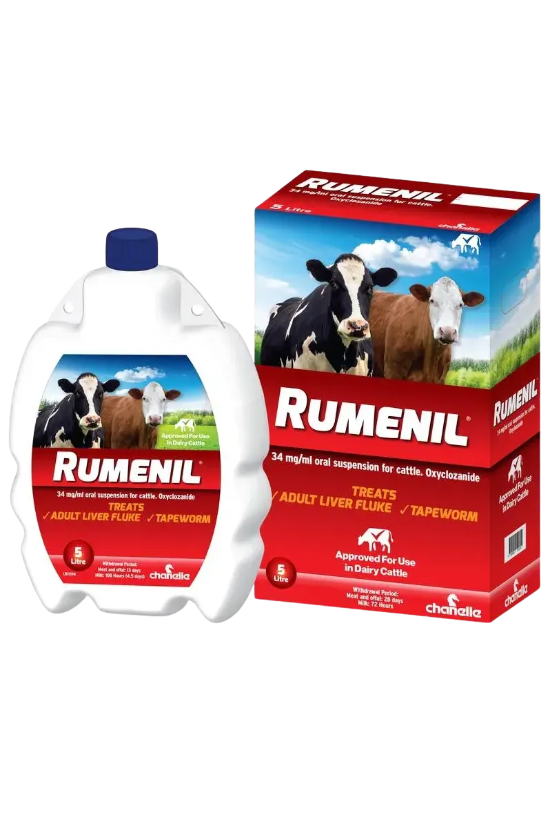
- Oxyclozanide
- Nitroxynil
- Closantel
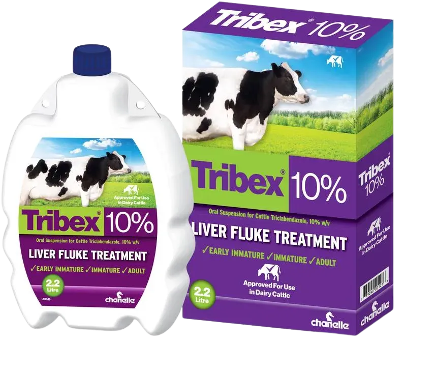
- Triclabendazole
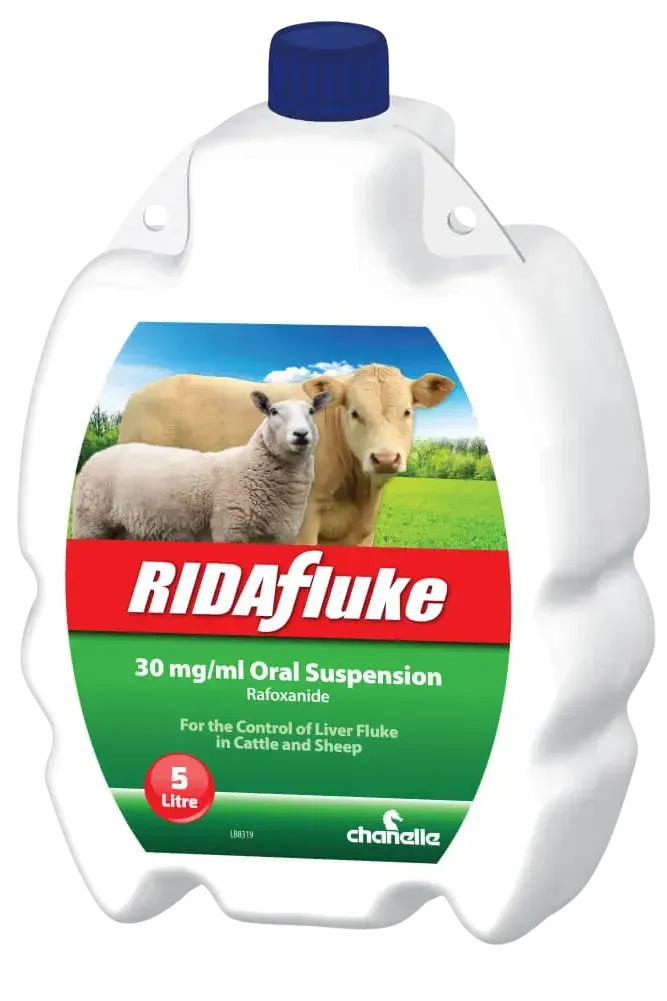
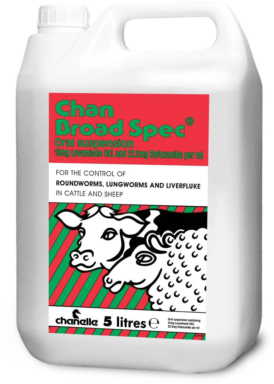
- Rafoxanide
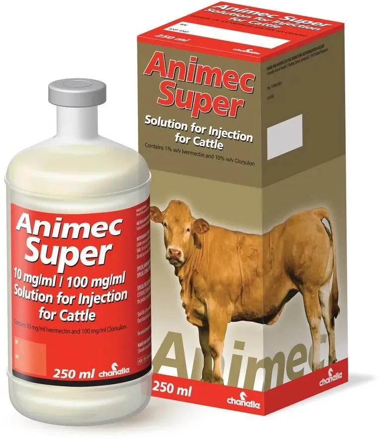
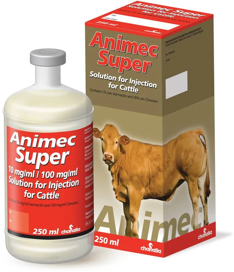
- Clorsulon
Different products target different stages of the parasite. Treatment choice should therefore be based on:
- Time of year
- Parasite stage present
- Resistance risk
Triclabendazole is the only active ingredient effective against all immature and adult stages of liver fluke. However, resistance has been reported in both the UK and Ireland, making responsible treatment use essential.
Sustainable Fluke Control
Key principles include:
Assess and Manage Grazing Risk
Map your farm
- Identify wet areas and mud snail habitats.
- Consider previous grazing history for both cattle and sheep.
Reduce exposure
- Fence off high-risk wet areas where possible.
- Choose alternative grazing where appropriate.
Use Diagnostics
- Test animals to determine whether infection is present.
- Use results to guide treatment timing.
Target Treatments
- Avoid unnecessary treatments to reduce resistance pressure.
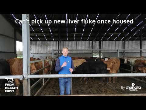
- Consider treatment when cattle are housed.
Work With Your Vet
- Veterinary advice can help develop a liver fluke control plan suited to your farm.
Rumen Fluke
Rumen fluke are common disease-causing parasites in tropical regions. In the UK and Ireland, the presence of rumen fluke (Calicophoron daubneyi) is increasingly recognised, and reports of rumen fluke eggs in faecal egg count samples have increased over the last 20 years.
The eggs closely resemble liver fluke eggs, so increased reporting may partly reflect improved identification techniques.
Their association with disease is less clear because clinical signs are often vague and overlap with liver fluke infection or other causes of poor thrive.
Adult rumen fluke living in the rumen are usually of low clinical significance, so the presence of rumen fluke eggs in faecal samples does not necessarily indicate that treatment is required.
However, heavy infestations of immature rumen fluke, which attach to the small intestine, can cause disease.
Severe infections may result in:
- Profuse watery diarrhoea (scour)
- Rapid weight loss and poor thrive
- Death in severe infections, particularly in youngstock
Because clinical signs are non-specific, rumen fluke infections can easily be overlooked or mistaken for other causes of scour.
Cases are most commonly reported during autumn and winter, although risk may continue beyond these seasons depending on environmental conditions.
It is important to remember that rumen fluke and liver fluke share the same intermediate host and have very similar lifecycles. As a result, where rumen fluke is present, liver fluke is often present too. In temperate regions, liver fluke remains the more significant disease threat.
How Infection Occurs
Rumen fluke shares a similar lifecycle to liver fluke, including the use of the mud snail as an intermediate host.
Diagnosis
Diagnosis can be challenging and should always consider both clinical signs and farm risk factors.
- Faecal egg counts detect eggs from adult rumen fluke only and are therefore not useful during early or acute disease. Eggs are absent during the damaging immature stages, so a negative faecal egg count does not rule out rumen fluke disease.
- Post-mortem examination is useful, particularly where large numbers of immature fluke are found in the small intestine.
Treatment
At present, no products are specifically licensed for rumen fluke in the UK.
However, research, including Animal Health Ireland guidance, indicates that oxyclozanide is effective against both immature and adult stages.
Treatment decisions should always be made in consultation with your vet and following an appropriate diagnosis.
Preventative management follows the same principles as liver fluke control.
Products





























Blog


.webp)







More Diseases
Know the solution.


CPD for SQPs & Vets
Sharpen your knowledge

DOsing Best Practices
Get the dose right every time