Gut Worms in Cattle
Quick Facts
Introduction
Gastrointestinal (GI) worms, often referred to as gut worms, stomach worms or roundworms, are an important group of parasites affecting grazing cattle in Ireland and the UK.
The disease they cause is known as parasitic gastroenteritis (PGE). It mainly affects youngstock during their first grazing season, although adult cattle can also experience production losses when worm burdens are high.
Cattle are typically affected by more than one species of worm at one time, so it is easier to discuss PGE in general, as symptoms, control and treatment usually overlap. Sometimes, however, we need a deeper understanding of specific disease caused by certain worm species. The two most important, ostertagiosis and cooperiosis, are discussed below.
Effective control of PGE relies on monitoring parasite risk, grazing management and responsible use of treatments.
The Impact of Gut Worms on Cattle Performance
Worm infections damage the digestive system and reduce the animal’s ability to utilise nutrients efficiently. As a result, infected cattle can show:
- Reduced appetite
- Poor growth rates
- Lower milk production
- Poor thrive
- Scouring
Importantly, the greatest losses often occur due to subclinical infections, where animals appear healthy but perform below expected levels.
PGE reduces growth rates by up to 30%, leading to a delay in:
- Reaching slaughter weight for beef cattle
- First service, or poorly grown pregnant heifers in dairy herds
Subclinical GI worm burdens can cause reduced yields of up to 1 kg per day in dairy cows.
How Infection Occurs
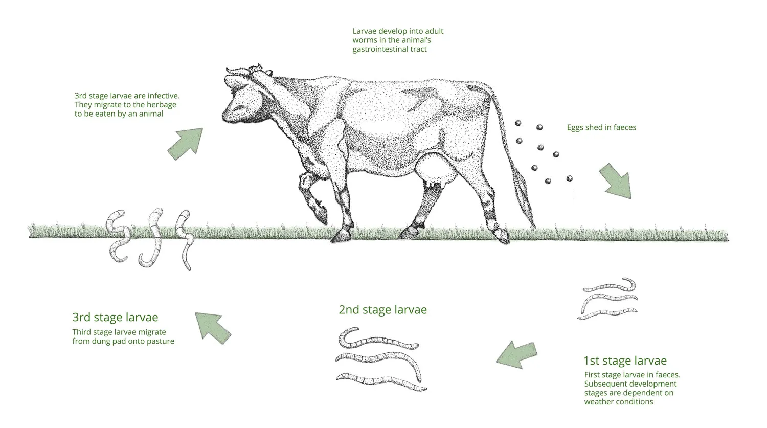
Infective larvae are present on pasture and are ingested while grazing. These larvae burrow into the gut wall and develop into adult worms. The adult worms produce eggs which are shed in dung, contaminating pasture and increasing infection pressure.
Without effective management, worm burdens can increase rapidly during the grazing season.
Eggs on the pasture develop best in mild and wet conditions, so a warm spring and summer with good rainfall can increase pasture contamination risk. Eggs can also remain viable over winter, seeding the pasture for the following grazing season.
The Main Gut Worms Affecting Cattle
The two most economically important gastrointestinal worms in cattle are Ostertagia ostertagi and Cooperia oncophora. Both are cattle-specific and have similar life cycles.
Cooperia oncophora
This worm infects the small intestine. It damages the intestinal lining and reduces nutrient absorption, leading to reduced weight gain and poorer feed efficiency.
Disease usually occurs as part of mixed PGE alongside ostertagiosis.
Acquired immunity is typically gained after the first grazing season.
Ostertagia ostertagi
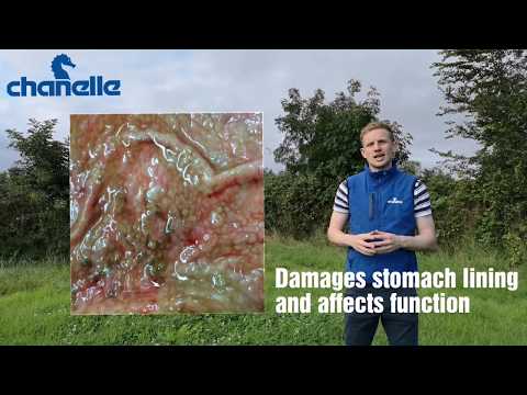
Also called the brown stomach worm, this parasite infects the abomasum, or true stomach. It damages the acid-producing glands of the abomasum, releasing acid which reduces digestive efficiency and suppresses appetite.
Acquired immunity typically takes two grazing seasons, and ongoing exposure is required to maintain it.
Disease caused by this parasite is known as ostertagiosis and may present in two ways.
Type I Disease
- This is the more straightforward presentation.
- The O. ostertagi burden increases in the abomasum during the grazing season as pasture contamination increases.
- It is often seen as a group of calves or cattle with profuse green scour, usually from August onwards.
Type II Disease
- Larvae ingested in late autumn enter a paused state called hypobiosis in the abomasum.
- During late winter or early spring, they continue their development.
- If there is a mass emergence, they can cause significant gut damage.
This may present as:
- Sudden death, even before scouring
- Sudden-onset scour
- Ill thrift and reduced appetite
In other animals, the emergence may be more gradual, without clinical signs of scour. If untreated, these animals can contribute to significant pasture contamination at turnout.
If Type II disease is suspected, speak to your vet. Blood sampling for the presence of pepsinogen, a digestive enzyme, can be useful.
Discuss treatment with your vet or RAMA. Strategic treatments over housing are useful in controlling Type II disease and reducing pasture burdens during the following season.
Why First-Season Grazers Are Most at Risk
Cattle gradually develop immunity to GI worms through exposure, usually over one to two grazing seasons. Calves entering their first grazing season have little immunity and are therefore most susceptible.
Risk timing varies depending on the production system:
- First-season suckler calves are usually at low risk until they are weaned. The highest-risk period is therefore late summer to autumn, and autumn- and winter-born calves are typically affected.
- First-season dairy calves are at risk from turnout, particularly when pasture contamination is high.
Older Animals
- Spring-born suckler calves are often more at risk during their second grazing season, as they are largely protected while on the cow.
- Immunity is acquired through exposure, so older animals are usually not clinically affected.
- Subclinical O. ostertagi infection can affect milk production.
- Discuss the benefits of targeted worming of dairy animals and the appropriate product with your vet.
Monitoring and Diagnosis
Effective control begins with monitoring parasite levels and animal performance.
Common diagnostic tools include:
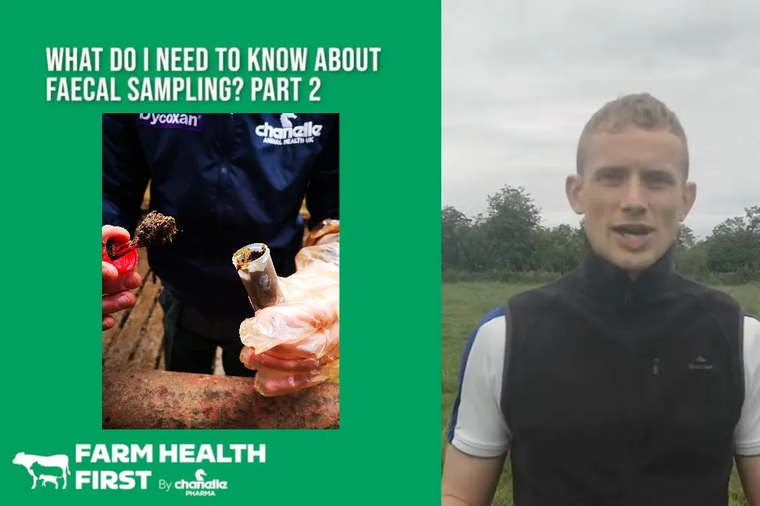
Faecal Egg Counts
- Can help identify the presence of GI worms and guide treatment decisions.
- Use group samples and discuss treatment if results exceed 200 eggs per gram.
Growth Monitoring
- Regular weighing of calves can identify animals with reduced daily liveweight gain, which may indicate parasite burdens.
Milk Antibody Testing
- Bulk milk tests measuring antibodies to Ostertagia can provide an indication of parasite exposure in dairy herds.
Post-mortem and Laboratory Testing
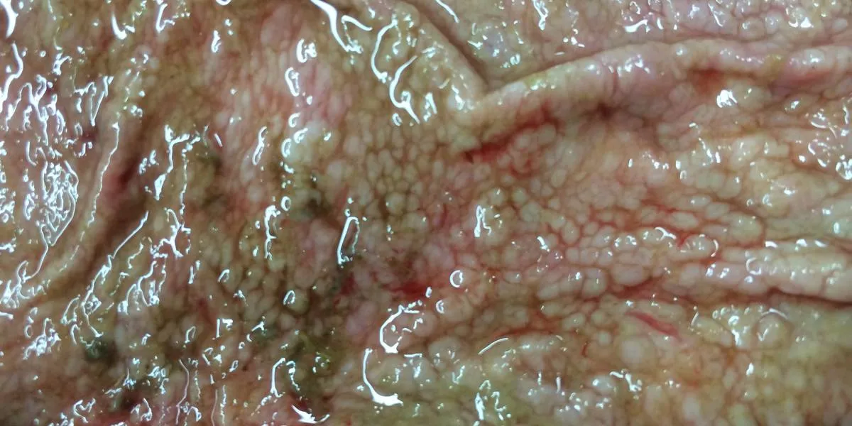
Sudden deaths are worth investigating, and post-mortem examinations can be very useful.
Control and Prevention of GI Worms
COWS and Animal Health Ireland recommend a sustainable, evidence-based approach to parasite control.
Key principles include:
Test Before Treating
- Use diagnostics and performance monitoring to assess whether treatment is needed.
- Routine blanket dosing should be avoided where possible.
Target Treatments
- Treat animals or groups at highest risk rather than the whole herd.
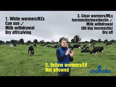
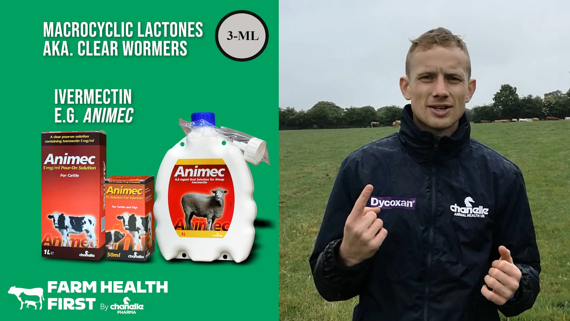
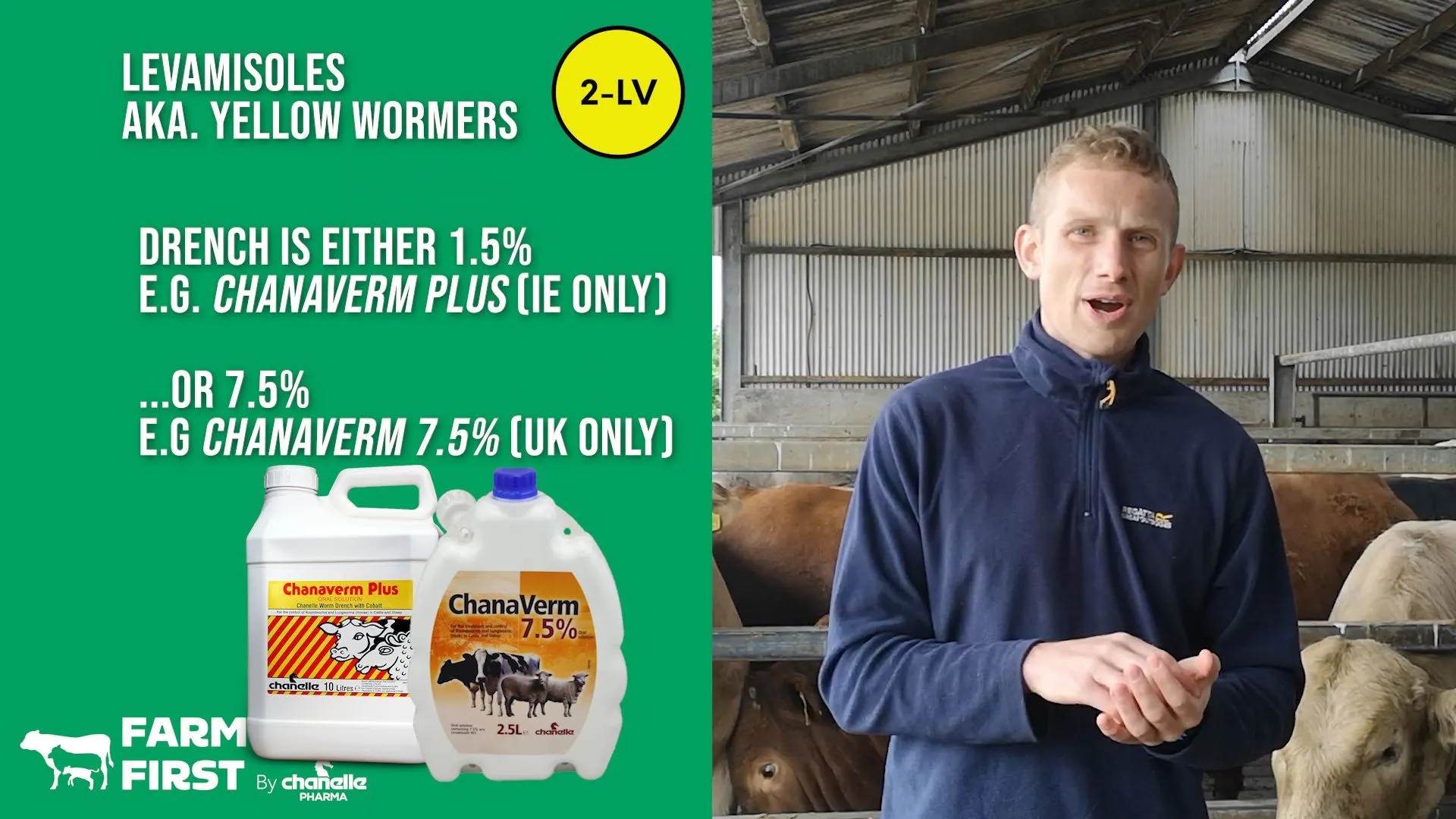
- Where treatments are used, rotate wormer groups to reduce resistance pressure.
Manage Pasture Contamination
- Grazing management can reduce parasite build-up and infection pressure.
- Co-grazing with sheep reduces pasture contamination, but be cautious of fluke.
- Use clean grazing, such as reseeded ground or silage aftermath, strategically.
- Consider rotating older cattle groups around the grazing block to reduce pasture contamination, but be aware of other disease risks such as Johne’s disease.
- Pasture mapping should form part of your control plan.
- Grazing history, weather, stocking density and rotation all influence larval survival and pasture risk.
Seek Veterinary Advice
- Work with your veterinarian, SQP or RAMA to develop a parasite-control strategy suited to your farm.
Responsible use of parasite treatments is essential to slow the development of anthelmintic resistance.
Tapeworms
Tapeworms have a different lifecycle from the roundworms that cause PGE.
Adult tapeworms are long, flat worms formed from chains of segments called proglottids, which contain eggs. These are shed in the dung and can be seen as small pieces of ribbon, often wriggling.
The eggs are released and ingested by intermediate hosts, where they mature and form infective cysts that are then picked up by the definitive host animal.
Cattle and sheep are the definitive hosts for Moniezia species. The intermediate host is a mite that lives on pasture.
Moniezia infection rarely causes clinical problems. Tapeworm is easily treated with most Group 1 wormers. Speak to your vet, SQP or RAMA for advice.
Cattle as the Intermediate Host
Cattle can act as the intermediate host for Taenia species. Cysts form in the muscle, leading to trimming and carcass rejection at slaughter. There is no treatment for this.
In the UK, sporadic outbreaks involving the human tapeworm Taenia saginata have occurred where it is suspected that animal feed was contaminated with human faeces.
Dog or fox tapeworms could also potentially cause cysts in cattle, although this is more commonly an issue in sheep.
Products





.webp)

















Blog






.webp)

.webp)


.webp)
.webp)





More Diseases
Know the solution.


CPD for SQPs & Vets
Sharpen your knowledge

DOsing Best Practices
Get the dose right every time